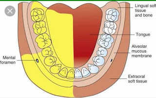
INFERIOR ALVEOLAR NERVE BLOCK
* Anatomical Landmarks of Inferior alveolar
nerve block:
• Mucobuccal fold
• Anterior border of Mandibular ramus
• External oblique ridge
• Internal oblique ridge
• Retromolar triangle
• Pterygomandibular ligament
• Buccal sucking pad
• Pterygomandibular space
* Nerves Anesthetized by Inferior alveolar
nerve block:
• Inferior Alveolar Nerve and its Sub-
divisions :
Mental Nerve
Incisive Nerve
Lingual Nerve
Buccinator Nerve
* Areas Anesthetized by Inferior alveolar
nerve block:
• Body of Mandible and an inferior portion of the ramus
• Mandibular teeth: Incisors, Canine,
Premolars and Molars
• Mucous membrane and underlying tissues
anterior to the first mandibular molar
(supplied by Lingual nerve)

* Note: To anesthetize the soft tissue
posterior to the 1st molar Long Buccal
nerve should be anesthetized.
* Amount of LA injected :- 1.8 ml
* Injection Technique used for Inferior
Alveolar Nerve Block (Patient Left Side):-
• The Patient is seated comfortably on
the Dental chair and the dentist stands on
the right side of the patient partially facing
the patient. For the Left side IANB the
Dentist stands more to the Front of the
patient. (These are for a Right Handed
Dentist)
• The Head of the patient is placed such that
the Body of Mandible is parallel to the
floor.
• The Dentist / Operator uses his/her left
index finger or Thumb to palpate the
Mucobuccal fold. If thumb is used the
index finger can be placed on extra orally
behind the ramus thus holding the
mandible between your thumb and index
finger to assess the width of the Ramus.
• The finger is then moved posterior to
contact the External oblique ridge on the
anterior border of the Ramus of mandible.
• From here it is moved up or Down in
search of the greatest depression called
the Coronoid Notch which is in direct line
with the Mandibular sulcus.
• The palpating finger is moved lingually
across the retromolar triangle and onto the
internal oblique ridge.
• The finger which is on the coronoid notch
and in contact with the internal oblique ridge
is moved to the buccal side to move the
Buccal Sucking pad away the path of
insertion.
• This gives better exposure to the internal
oblique ridge, the pterygomandibular
raphae and the pterygomandibular
depression.
• The Needle is inserted parallel to the
occlusal plane of the mandibular teeth from
the opposite side (Approximately over
opposite side Premolar) of the mouth at a
level bisecting the finger or thumbnail
penetrating the tissues of the
pterygotemporal depression, and entering
the pterygomandibular space.
• The needle is penetrated until bone is
contacted on the internal surface of the
ramus of the mandible.
• After contacting bone the needle is
withdrawn about 1mm and then 1 to 2 ml of
solution is deposited slowly over a duration
of 2 minutes into the tissue.
* Technique for anesthetizing lingual nerve :
• Needle is withdrown about half of its length
which was inserted and local anesthetic
Solution is injected at the area which
anesthetized lingual nerve.
* The signs and symptoms of an inferior alveolar block
are:
• Tingling and numbness of the lower lip (however it is not
an indication of depth of anesthesia).
Tingling and numbness of the tongue (see Lingual Nerve Block). No pain is felt during dental treatment.


Comments
Post a Comment