INFRAORBITAL NERVE BLOCK

• Infraorbital Ridge
• Infraorbital depression
• Supraorbital notch
• Infraorbital notch
• Anterior teeth
• Pupils of the eyes
* Nerves Anesthetized:
• Infraorbital nerve
• Anterior Superior Alveolar nerve
• Middle Superior Alveolar nerve
• Inferior Palpebral Nerve
• Lateral Nasal Nerve
• Superior Labial Nerve
* Areas Anesthetized:
• Incisors
• Cuspids
• Bicuspids
• Mesiobuccal root of first molar on the side affected
• Bony Support and Soft tissue
• Upper lip
• Lower eyelid
• Portion of the nose on the same side

* Technique for infra orbital nerve block :-
√ Needle Pathway during Insertion:
There are 2 approaches to the Infra orbital nerve block, the most commonly used being Bicuspid approach and Central incisor approach.
1. Bicuspid Approach: Needle is inserted into the mucosa and the areolar tissue using the Maxillary Bicuspid as the guide and the needle should pass beneath and lateral to the external maxillary artery and the anterior facial vein.
2. Central Incisor Approach: Needle is inserted into the mucosa and the alveolar tissue using the Maxillary Central Incisor as the guide and it passes beneath the angular head of the Quadratus labii superiors muscle. It proceeds anterior to the origin of the caninus muscle and beneath the external maxillary artery and the anterior facial vein.
√ Injection Technique:
* Operator position :-
• For the right side, the dentist stands on the right side of the chair partially facing the patient.
• For the left side the operator stands more to the front of the Patient.
* Patient position :-
Patient should be placed in a comfortable position in the chair such that the maxillary occlusal plane is at a 45 degree angle to the floor.
- The infraorbital and supraorbital notches are palpated while the patient is asked to look forward.
- Draw an imaginary Straight line through the following landmarks:
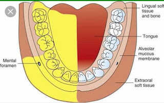
Mental Foramen,
Bicuspid Teeth,
Infraorbital foramen

- Locate the infra orbital notch with your Thumb finger and after palpating the finger should be moved downwards about 0.5 cm until a shallow depression is felt.
- Use the index finger to retract the Lip exposing the muco buccal fold.
- The needle should be inserted at this location at a sufficient distance (5mm) from the labial plate to pass over the canine fossa.
- The Needle after insertion in the desired location should be guided with the help of the thumb which was placed on the infraorbital foramen.
- The needle should be guided into position so that it contacts the bone at the entrance to the foramen.
- Make sure that the needle does not penetrate more than 3/4th Inch and use the thumb to prevent the needle from entering the orbital cavity.
- 1.5 ml of Local anesthetic solution is deposited and Thumb is used to hold the needle in position till the end.
* Symptoms of Anesthesia:
• Objective Symptoms: Instrumentationshould be done, use either the Tip of a blunt Probe or Elevator to check for Objective symptoms.The anesthetized tissue and teeth should be pressed and tapped as compared to the other un-anesthetized site.
• Subjective Symptoms: Tingling and numbness of the upper lip, lower eyelid and side of the nose on the side affected will always be present but are not necessarily an indication of good anesthesia.
Comments
Post a Comment